
T. brevicauda Boulenger, 1916

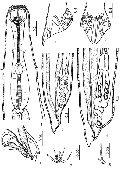
Figures 1-8:
1. Esophageal region, ventral view. 2. Male tail, lateral view. 3. Male tail, dorsoventral view. 4-5. Tail of female. 6. Genital cone, lateral view. 7. Tip of genital cone, ventral view. 8. Fused spicule tips of male.

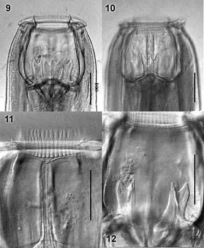
Figures 9-12
9. Buccal capsule, dorsoventral view. Arrow marks amphid or lateral papilla 10. Buccal capsule, lateral view. 11. Mouth collar, lateral view, showing 2 submedian papillae, each with a short bilobed process on the stalk (horizontal arrows) and a long slender tip, and an amphid (vertical arrow).
© (contents) R.J.
Lichtenfels, V.A. Kharchenko,
G.M. Dvojnos 2003
Design and programming: Yuriy Kuzmin,
2003