
P. skrjabini (Ershov, 1930) Ershov, 1943

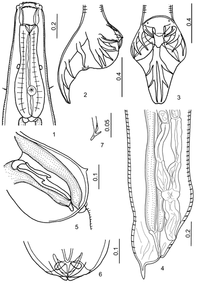
Figures 1-7:
1. Esophageal region, ventral view. 2. Male tail, lateral view. 3. Male tail, dorsoventral view. 4. Tail of female. 5. Genital cone, lateral view. 6. Tip of genital cone, ventral view. 7. Fused spicule tips of male.

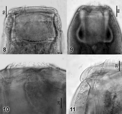
Figures 8-11:
8. Buccal capsule, dorsoventral view. 9. Buccal capsule, lateral view. 10. Submedian papillae, elements of ILC and ELC, and cuticular shelf of lining of BC. 11. Submedian papillae, elements of ILC, and dorsal gutter (arrow).
© (contents) R.J.
Lichtenfels, V.A. Kharchenko,
G.M. Dvojnos 2003
Design and programming: Yuriy Kuzmin,
2003