
Cylicocyclus adersi (Boulenger, 1920)
Ershov, 1943

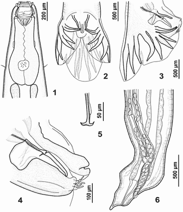
Figures 1-6. Cylicocyclus adersi, drawings. 1 - esophageal region, ventral view; 2 - male tail, ventral view; 3 - male tail, lateral view; 4 - genital cone, lateral view; 5 - fused spicule tips of male; 6 - tail of female.

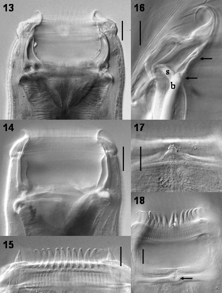
Figures 13-20. Cylicocyclus adersi, photomicrographs. 13 – anterior extremity, dorsoventral view, showing walls of buccal capsule; 14 - anterior extremity, lateral view; 15 – anterior extremity, dorsoventral view, showing dorsal gutter and elements of ILC and ELC; 16 – dorsal segment of esophageal funnel, dorsoventral view, showing protrusion in esophageal funnel; 17 – the structure of mouth collar, lateral view, showing ELC, pulp of ELC, septum intracoronare, ILC and support of ELC; 18 - amphid; 19 – submediam papilla; 20 – elements of external and internal radial crowns.
© (contents) R.J.
Lichtenfels, V.A. Kharchenko,
G.M. Dvojnos 2003
Design and programming: Yuriy Kuzmin,
2003