
C. montgomeryi (Boulenger, 1920) K'ung, 1964

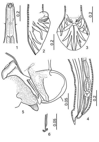
Figures 1-6:
1. Esophageal region, ventral view. 2. Male tail, lateral view. 3. Male tail, dorsoventral view. 4. Tail of female. 5. Genital cone, lateral view. 6. Fused spicule tips of male.

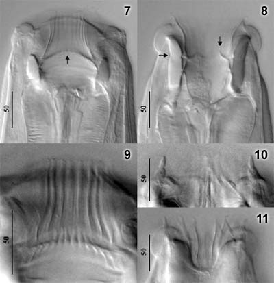
Figures 7-11:
7. Buccal capsule, dorsoventral view, showing insertion line fo elements of ILC and its junction with the ELC (arrow). 8. Buccal capsule, lateral view. Vertical arrow marks anterior tip of ILC and horizontal arrow marks junction of wall of BC and large support for ELC. 9. Elements of ELC and ILC. 10. Submedian papillae. 11. Wall of BC, lateral view, showing large gap in the large support for the ELC.
© (contents) R.J.
Lichtenfels, V.A. Kharchenko,
G.M. Dvojnos 2003
Design and programming: Yuriy Kuzmin,
2003