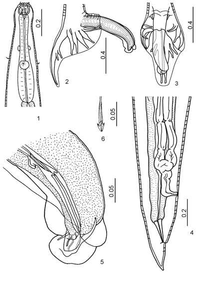

B. ivashkini Tshoijo in Popova, 1958

Figures 1-6:
1. Esophageal region, ventral view. 2. Male tail, lateral view. 3. Male tail, dorsoventral view. 4. Tail of female. 5. Genital cone, lateral view. 6. Fused spicule tips of male.

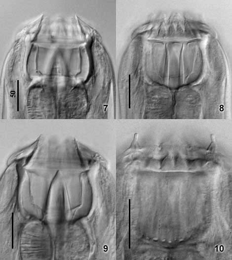
Figures 7-10:
7. Buccal capsule, dorsoventral view. 8. Buccal capsule, ventral view, showing 2 long, slender subventral esophageal teeth, and elements of ILC and ELC. 9. Buccal capsule, dorsal view, showing large dorsal esophageal tooth. 10. Submedian papillae and elements of ILC and ELC.
© (contents) R.J.
Lichtenfels, V.A. Kharchenko,
G.M. Dvojnos 2003
Design and programming: Yuriy Kuzmin,
2003