
Leonid Frantsevich
Experimental evidence on actuation
and performance of the elytron-to-body articulation in a diving
beetle, Cybister laterimarginalis (Coleoptera, Dytiscidae).
Journal of Insect Physiology,
2012, 58: 1650Ц1662.
Doi: http://dx.doi.org/10.1016/j.jinsphys.2012.10.006.
Movements of the elytra and axillary sclerites
were video recorded in tethered flying beetles and in manipulated
mounts of C. laterimarginalis, Dytiscus dimidiatus, and Acilius
sulcatus. Mesothoracic axillary plates are homologues of those
in the metathorax. Two anterior axillaries (Ax1, Ax2) fuse together;
they are hinged to the third axillary plate (Ax3). In turn, Ax3
is hinged with the elytron. During take-off, a beetle abducts
and highly elevates its elytra (1), then droops the elytra in
a flatly spread position (2); closing is adduction in a horizontal
plane (3). These steps have been simulated: (1) by pressing down
the anterior horn; (2) occurred spontaneously after release of
pressing; (3) the elytron closed flatly either by manual turn
of the elytron or by manual elevation of the prothorax. Anterior
axillaries rotated forward and down during (1), returned during
(2) and remained immobile during closing. Ax3 is folded between
the closed elytron and Ax1+Ax2; it unfolds during opening. Two
hinges of Ax3 form a Z-configuration and provide a linked drive
for complicated rotation of the elytron. Opening was impaired
in vivo if tergal leg protractor and depressor were disabled,
closing did not suffer. Closing was prevented by excision in the
hind edge of the pronotum, not harmful for opening. Role of direct
and indirect muscles in transient elytral movements is discussed.
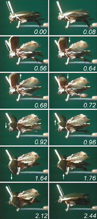 |
Opening and closing of elytra in a tethered Cybister laterimarginalis.
Selected frames from a video film, time in italics. Neighbour
frames in rows. A straw rod glued normally to the prothorax indicates
its orientation relative to the fixed pterothorax. The initial
orientation is indicated with a thin white line in (0.00 s) and
copied in (0.56 s), indicating depression of the prothorax during
opening. Elevation of the prothorax during closing is shown in
(2.12 and 2.44 s) in the same fashion. Alternating elevations
and depressions of the spread elytra are indicated with arrows
in serial frames. |
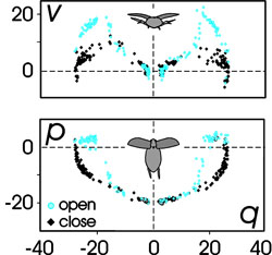
Trajectory of apical marks on the elytra in
Cybister laterimarginalis during opening and closing. Top panel
Ц frontal body-fixed plane, bottom panel Ц equatorial plane. Tip
of the scutellum is the origin of body-fixed reference. Scales
in mm. Elytra are elevated higher during opening.
Reconstruction of the structure and performance of the elytral
articulation must explain peculiarities of elytral movements in
diving beetles. |
 |
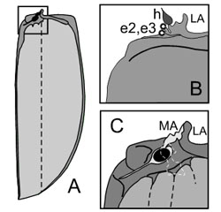
|
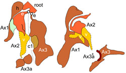
|
(A) Elytron in C. laterimarginalis, ventral
face. Articulatory root area in a box.
(B) Root area, dorsal face, (C) ventral face.
Circles e2, e3 Ц articulations with elytral
processes of axillaries 2 and 3, h Ц head of Ax1, LA, MA Ц lateral
and medial apophyses of the root. |
Homology between elytral and wing axillary
plates Ax1-Ax3a. Abbreviations: c1 Ц crease between Ax2 and Ax3;
e Ц elytral processes to the root; v Ц ventral subplate of Ax2. |
(click on the image to view movie
in separate window)
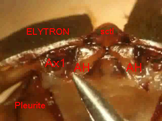
Microfilming of dead mount of C. laterimarginalis.
Front view at the right elytron and mesothorax. (sctl Ц scutellum)
Manipulated opening of the right elytron by
pressing onto the anterior horn (AH) simulates contraction of
the direct tergo-pleural muscle M33 and indirect leg protractor
and depressor. Anterior axillaries Ax1, Ax2 rotate together with
the elytron, the elytron elevates. The open elytron falls down.
Manipulated closing was achieved by turn of the elytral blade
with a forceps. Anterior axillaries remain immobile.
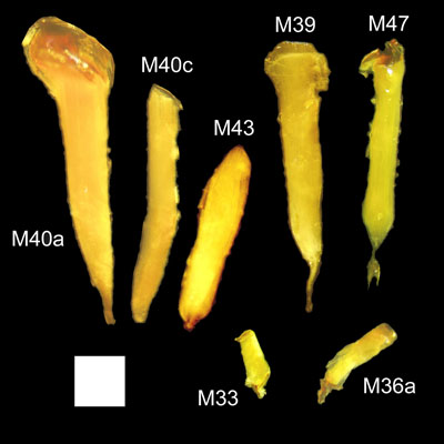
Direct and indirect mesothoracic
wing muscles in Cybister laterimarginalis. Whole mounts in glycerol,
dark field photographs. Muscle numbers by Larsen (1946). Scale
square: 1 mm2.
M40a, c Ц leg retractors.
M33 Ц direct opener of the elytron.
M36a Ц direct closer ?
Muscles of dual function:
M39 Ц leg protractor, opener.
M47 Ц leg depressor, opener.
M39 Ц leg retractor, closer.
(click on the image to view movie
in separate window)
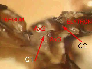
Microfilming of dead mount of
C. laterimarginalis. Right side, rear view.
Rotation of Ax3 upon manipulated
closing and opening by the blade of the elytron. Creases C1 and
C2 fold or unfold. Anterior axillaries Ax1, Ax2 remain immobile.
Double rotation about two creases provides a linked drive with
the single degree of freedom.
(click on the image to view movie
in separate window)
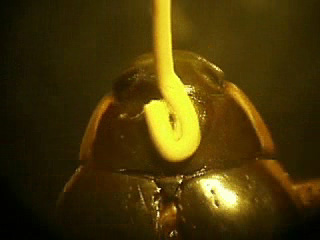
Manipulated closing by the prothorax
in C. laterimarginalis simulates contraction of muscles
elevating the head and prothorax. The posterior edge of the pronotum
acts on the root of the elytron.
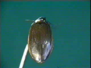
(click on the image to view movie
in separate window)
High opening, flutter, and flat
closing in Dytiscus dimidiatus.
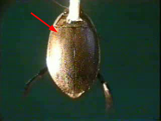
(click on the image to view movie
in separate window)
Abortive opening and normal closing
of the left elytron in Acilius sulcatus after excision of the
left midleg and inactivation of indirect leg protractor and depressor.
|
