Keys to genera
1 (10) Buccal capsule small, not wider than anterior part of
oesophagus, or absent.
2 (3) Cuticular spines present on surface of body in anterior
part (Fig. 1). Parasitic in Scincidae (Reptilia: Sauria) in Australia.
– Pneumonema Johnston, 1916.

Fig. 1. Anterior part of P.
tiliquae body, SEM
3 (2) Surface of body without spines.
4 (5) Surface of body cuticle with longitudinal crests (Fig.
2). Buccal capsule absent. Three onchia present on oesophageal
apex. Parasitic in mouth cavity of Ameiva ameiva (Reptilia:
Sauria: Teiidae). – Chabirenia
Lhermitte-Vallarino, Bain, Deharo et al., 2005.
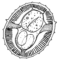
Fig. 2. Cross-section through
the C. cayennensis mid-body showing longitudinal crests
(after Lhermitte-Vallarino et al., 2005).
5 (4) Surface of body cuticle without longitudinal crests.
6 (7) Body cuticle inflated, at least on some parts of body
(Fig. 3). Buccal capsule present, in some species consisting of
anterior and posterior segments. Parasitic in amphibians (Anura,
Caudata, Gymnophiona) and lizards from Iguania (Agamidae, Chamaeleonidae,
Iguanidae); probably, present in some Gekkonidae. – Rhabdias
Stiles et Hassall, 1905.
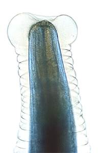
Fig. 3. Anterior part of R.
sphaerocephala showing typical cuticular inflation in a live
specimen.
7 (6) Body cuticle not inflated, just thickened in anterior
and posterior parts, often transversely striated. Parasitic in
snakes (Reptilia: Serpentes).
8 (9) Apical extremity with 8–10 conical projections directed
anteriorly (Fig. 4) – Acanthorhabdias
Pereira, 1927.
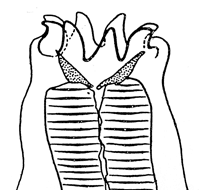
Fig. 4. Anterior end of A.
acanthorhabdias (after Pereira, 1927).
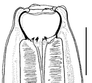
9 (8) Apical extremity with 6 small, equal or sub-equal lips
arranged in lateral groups (Fig. 5). Buccal capsule funnel-shaped,
or absent in some species. – Serpentirhabdias
Tkach, Kuzmin et Snyder, 2014.

Fig. 5. Apical extremity of
S. eustreptos (SEM) showing lips arranged in lateral
groups.
10 (1) Buccal capsule large, thick-walled, wider than anterior
end of oesophagus.
11 (12) Six onchia present on oesophageal apex (bottom of
buccal capsule) (Fig. ). Parasitic in Anguidae (Reptilia: Sauria).
– Entomelas Travassos, 1930.
 A A 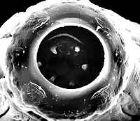 B B
Fig. 6. Entomelas
ophisauri: lateral view (A) and sub-apical
view (B) of anterior end showing large buccal
capsule and onchia.
12 (11) Onchia absent. Parasitic in Scincidae (Reptilia: Sauria).
13 (14) Apical extremity globular, with dorsal and ventral
pseudolabia (Fig. ). – Neoentomelas
Hasegawa, 1989.
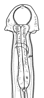
Fig. 7. Anterior part of N.
asatoi body (lateral view) showing characteristic shape of
anterior end.
14 (13) Apical extremity with 6 small lips, or with 4 submedian
lips and 2 lateral pseudolabia (Fig.8 ). – Kurilonema
Szczerbak et Sharpilo, 1969.
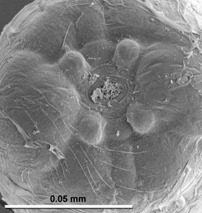
Fig. 8. Apical extremity of
K. browni showing submedian lips and lateral pseudolabia.
|